
Snapping Scapula Syndrome: Causes, Symptoms & Treatment
The “Rhomboid Strain” Impostor
Pain along the medial border of the scapula is often attributed to rhomboid strain, poor posture, or thoracic spine dysfunction. But when this pain is accompanied by reproducible grinding, popping, snapping, or clunking during scapular motion, the problem may lie elsewhere: at the scapulothoracic interface.
This is Snapping Scapula Syndrome (SSS) an uncommon but frequently overlooked cause of persistent periscapular pain and scapulothoracic bursitis.
What Is Snapping Scapula Syndrome?
Snapping Scapula Syndrome is characterized by painful mechanical crepitus arising from the scapulothoracic articulation. The crepitus may be audible, palpable, or both and is typically reproduced during active scapular movement.
The scapulothoracic articulation is not a true synovial joint. It is a functional articulation in which the concave anterior surface of the scapula glides over the convex thoracic cage, with muscles and bursae creating a low-friction interface between the two surfaces.
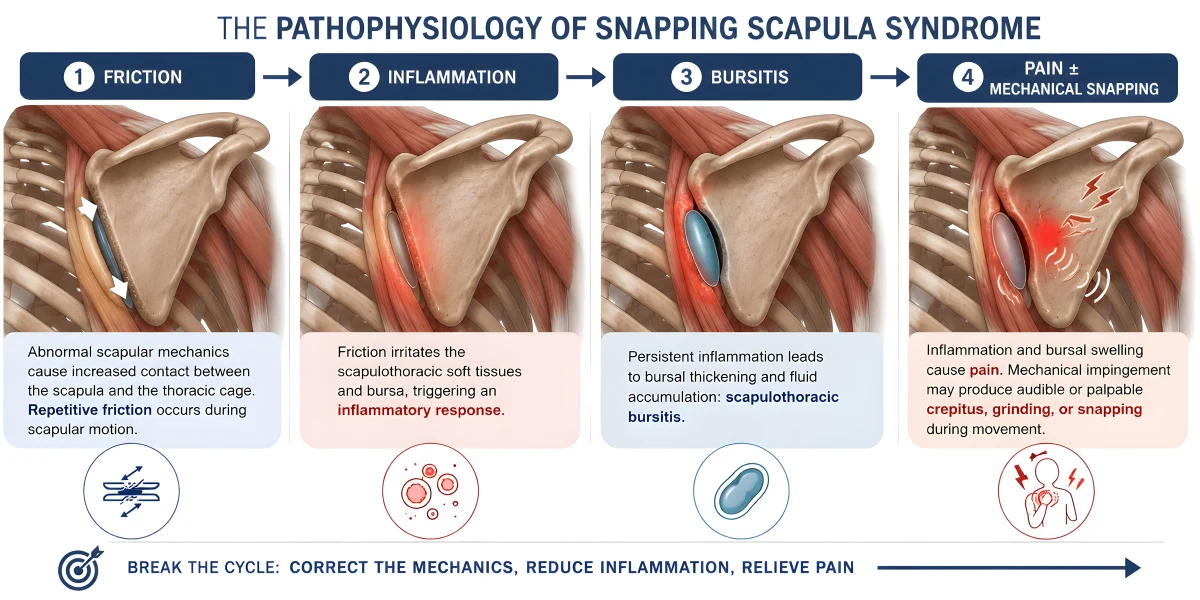
When this smooth gliding mechanism is disrupted, repetitive contact between the scapula, soft tissues, and ribs can generate:
Friction → inflammation → bursitis → pain ± mechanical snapping
The underlying abnormality may be functional, soft-tissue, osseous, or a combination of these factors.
 Pathophysiology
1. Scapulothoracic Bursitis
Repeated mechanical friction can irritate the scapulothoracic bursae, particularly around the superomedial scapular border.
Chronic irritation leads to inflammation, bursal thickening, and pain, which may further alter normal scapular mechanics and perpetuate the problem.
2. Scapular Dyskinesis & Muscular Imbalance
Abnormal scapular positioning or movement changes the relationship between the scapula and thoracic cage.
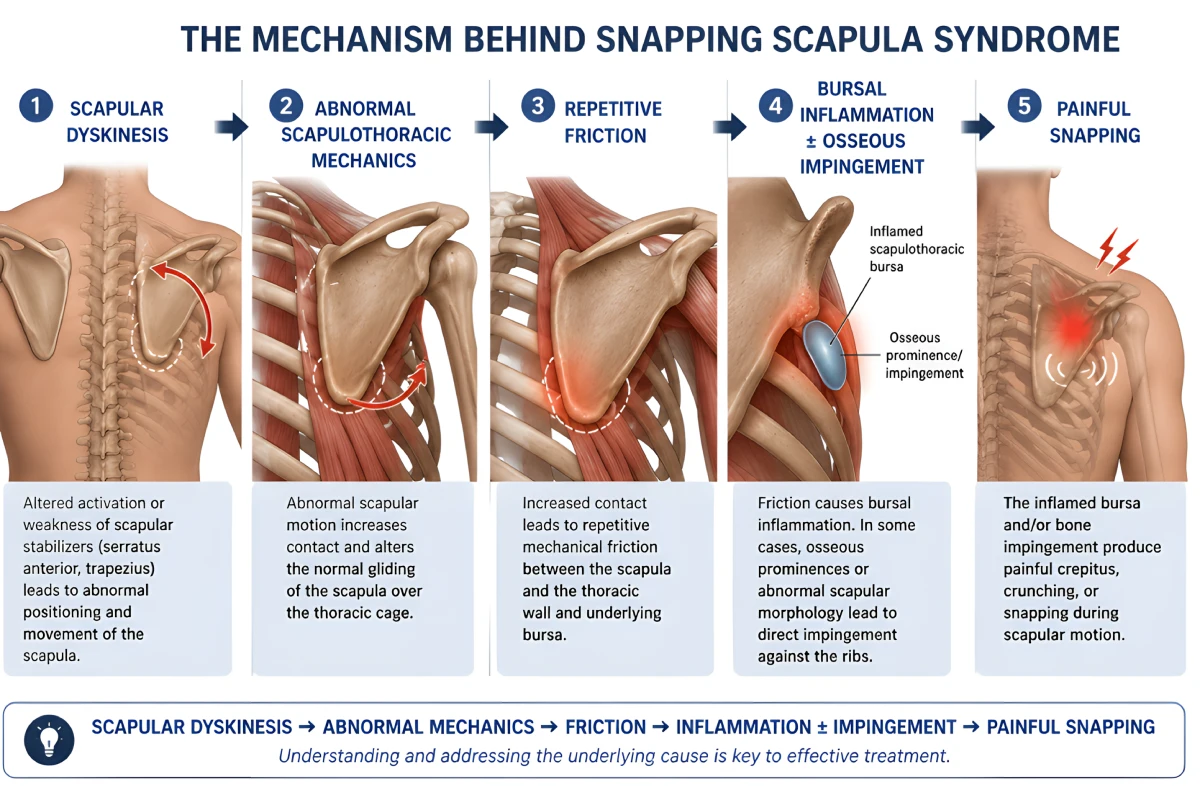
Poor neuromuscular control or weakness of the serratus anterior and trapezius, combined with abnormal tension or imbalance of the levator scapulae and rhomboids, may increase scapulothoracic contact and friction.
This can create a self-perpetuating mechanical cycle:
Scapular dyskinesis → abnormal mechanics → increased contact → repetitive friction → inflammation → pain → further dyskinesis
3. Osseous Abnormalities
Some patients have a structural cause producing direct mechanical impingement against the thoracic cage.
Potential abnormalities include:
- Prominent superomedial scapular angle
- Abnormal scapular morphology
- Luschka tubercle
- Osteochondroma
- Malunited scapular or rib fractures
- Rib deformities
- Other bony prominences involving the ventral scapular surface
In these cases, the snapping may have a particularly distinct mechanical component.
Typical Clinical Presentation
Patients commonly describe deep, poorly localized pain around the medial or superomedial border of the scapula.
Characteristic features include:
- Painful palpable or audible crepitus
- Grinding, popping, snapping, or clunking
- Symptoms reproduced during scapular elevation, retraction, rotation, or circumduction
- Pain aggravated by repetitive overhead activity
- Symptoms during throwing, reaching, pushing, or lifting
- Localized tenderness along the medial or superomedial scapular border
- Associated scapular dyskinesis in some patients
An important distinction
Scapulothoracic crepitus does not necessarily mean disease.
Painless crepitus can occur in otherwise asymptomatic individuals. The finding becomes clinically significant when the mechanical phenomenon is associated with pain, functional impairment, and reproducible symptoms.
Clinical Examination
The examination should evaluate the entire shoulder girdle and kinetic chain, rather than simply focusing on the painful rhomboid region.
Inspection
Observe scapular position at rest and during active arm elevation and lowering.
Look for:
- Scapular dyskinesis
- Winging
- Abnormal upward or downward rotation
- Excessive anterior tilt
- Asymmetric scapular movement
- Abnormal medial-border prominence
Dynamic Palpation
Palpate the medial and superomedial scapular borders while the patient actively moves the shoulder.
The objective is to determine whether the patient’s characteristic pain and crepitus can be reproduced mechanically.
Do Not Forget the Differential Diagnosis
A complete examination should also include:
Cervical spine → neurologic examination → glenohumeral joint → AC joint → scapulothoracic articulation
This is essential because scapular symptoms may be secondary to pathology elsewhere in the shoulder girdle.
Imaging: Match the Modality to the Suspected Cause
Radiographs
Useful as an initial assessment for obvious osseous abnormalities but may fail to demonstrate subtle abnormalities of the scapulothoracic interface.
CT / 3D CT
Particularly valuable when a bony mechanical cause is suspected.
3D reconstruction can define scapular morphology and demonstrate:
- Superomedial prominence
- Luschka tubercle
- Osteochondroma
- Post-traumatic deformity
- Abnormal scapula–rib relationships
MRI
Best suited to the evaluation of soft-tissue pathology, including:
- Scapulothoracic bursitis
- Bursal fluid
- Soft-tissue masses
- Muscle abnormalities
- Associated shoulder pathology
Dynamic Ultrasound
Provides real-time assessment of the symptomatic region and can identify bursal pathology.
It also offers an important practical advantage: ultrasound-guided diagnostic and therapeutic injection.
Why Is Snapping Scapula Frequently Misdiagnosed?
Because medial scapular pain is nonspecific.
SSS may mimic:
- Rhomboid strain
- Levator scapulae pain
- Myofascial pain
- Cervical radiculopathy
- Thoracic spine pathology
- Scapular dyskinesis
- Glenohumeral pathology with secondary dyskinesis
- Long thoracic nerve dysfunction
- Spinal accessory nerve dysfunction
Therefore:
The sound alone is not the diagnosis. The combination of pain, reproducible mechanical symptoms, abnormal scapular mechanics, and appropriate imaging establishes the clinical picture.
Treatment
First Line: Treat the Mechanics
Most patients should initially be managed non-operatively.
The objective is not simply to reduce pain. It is to normalize the biomechanics responsible for abnormal scapulothoracic contact.
Rehabilitation should emphasize:
- Scapular motor control
- Serratus anterior strengthening
- Middle and lower trapezius strengthening
- Correction of scapular dyskinesis
- Restoration of thoracic mobility
- Postural correction when appropriate
- Correction of muscular imbalance
- Treatment of associated glenohumeral pathology
- Progressive return to overhead activity
Activity modification and appropriate analgesic or anti-inflammatory measures can be useful during symptomatic phases.
Diagnostic & Therapeutic Injection
When scapulothoracic bursitis is suspected, injection of the symptomatic bursa with local anesthetic ± corticosteroid may serve two purposes.
A significant temporary reduction in pain supports the scapulothoracic bursa as an important pain generator, while corticosteroid may provide therapeutic relief.
Ultrasound guidance improves localization and injection accuracy.
When Should Surgery Be Considered?
Surgery should be reserved for persistent, function-limiting symptoms despite an adequate period of structured rehabilitation, particularly when a reproducible mechanical source has been identified clinically and/or radiologically.
The surgical strategy should be directed toward the underlying pathology.
Arthroscopic Scapulothoracic Bursectomy
Indicated primarily for persistent symptomatic scapulothoracic bursitis that has failed conservative treatment.
Superomedial Scapuloplasty / Partial Scapulectomy
Bone resection may be added when imaging and clinical findings demonstrate a relevant osseous prominence responsible for mechanical impingement.
The principle is important:
Do not treat every snapping scapula with the same operation.
Surgery should be pathology-driven rather than routinely combining bursectomy, muscle release, and bone resection.
Clinical Takeaway
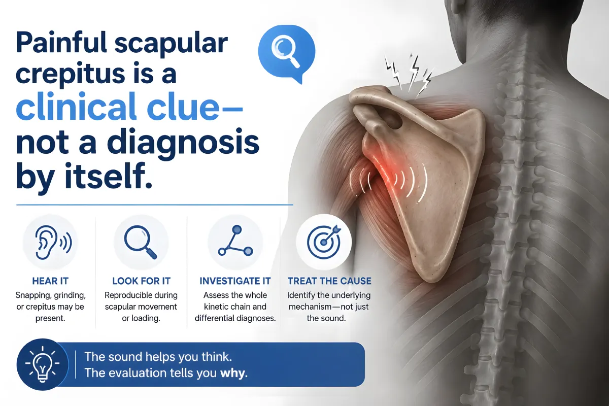
Painful scapular crepitus is a clinical clue not a diagnosis by itself.
When persistent medial or superomedial scapular pain is associated with reproducible snapping, grinding, or crepitus, consider Snapping Scapula Syndrome before labelling the problem as another rhomboid strain.
The key is to determine why the scapula is snapping.
The mechanical pathway
Scapular dyskinesis
↓
Abnormal scapulothoracic mechanics
↓
Increased scapula–thoracic contact
↓
Repetitive friction
↓
Scapulothoracic bursal inflammation ± osseous impingement
↓
Painful crepitus / snapping
Treat the cause, not simply the sound.
Most patients should begin with targeted biomechanical rehabilitation. In carefully selected patients with persistent symptoms and a clearly defined mechanical pathology, arthroscopic scapulothoracic surgery can provide effective treatment.